reflections
3:40 AM
time flies. and now, this will be my last entry for the Biology Journal project. i can say that it has been a pleasant and enriching experience to learn biology in this way. and the fact is that, i have learnt a great deal on the 2 topics that i have researched on- digestion and transport in living things.
although it has been strenuous for me, trying to keep up with the constant updates of my entries, i found out that i can learn a lot more just by researching on the topics that i chose and locating key words or important details from the chunk of information i received.
i admit it was not an easy task to make a good entry, but at least i tried my best to do the best out of it. and although this will be my last post, the knowledge that i have learnt through everything i have done will always remain as mine.
transport system[10]
8:11 AM
After learning such a great deal about the transport system in plants, let me add on to some other findings on xylem and phloem tubes:
xylem:
- made of dead cells
- thick cell walls
- cell walls made of lignin (a hard cellulose-like substance which gives rigidity to plant tissues)
- impermeable
- cross walls absent
- no cytoplasm
- transport of water and minerals
- carried to leaves
- upward flow
- tissue has fibres
phloem:
- made of living cells
- thin cell walls
- cell walls made of cellulose
- permeable
- has perforated cross walls called sieve plates
- cells lined with cytoplasm strands
- transport of food
- carried to growing parts and storage organs
- upward and downward flow
- tissue has companion cells
Ok, this marks the end of my biology journal. i hope that all of you will take away some valuable knowledge with you after reading my entries. thank you(:
transport system[9]
7:15 AM
Sugar are made in the leaves using the water transported by the xylem vessels and the carbon dioxide which diffuses into the leaves from the air. These sugar are carried to every other part of the plant that needs them, such as the roots, buds and younger leaves, by the phloem tubes. The phloem tubes also carry other substances made by the plant cells such as the amino acids.
Unlike xylem (which is composed primarily of dead cells), the phloem is composed of still-living cells that transport sap. The sap is a water-based solution, but rich in sugars made by the photosynthetic areas. The phloem tube is made of cellulose and perforated end walls called sieve plates. It is permeable, and are surrounded by companion cells.
Together xylem and phloem form the vascular tissue, often also referred to as the vascular bundle. The two types of vessel are always found together, but they occupy slightly different locations in the root and the stem.

transport system[8]
6:30 AM
Xylem vessels are like long drainpipes reaching all the way from the roots to the tip of every leaf. They are made up of hollow dead cells joined end to end.
Water and dissolved mineral salts are drawn up the xylem vessels when water from the leaves evaporates and diffuses out through tiny holes called stmata into the air. This process whereby plants lose water due to evaporation is called transpiration. As transpiration continues, more water is pulled up the xylem as though someone is "sucking" water up the plant. The movement of water with dissolved mineral salts through the plants is called the transpirational pull. Other phenomena that cause xylem sap to flow are the following- cohesion and adhesion & root pressure.
Transpirational pull- As water evaporates from the leaves of the plant,there is a suction created by this action resulting in a pull drom the roots..
Cohesion and Adhesion- The water molecules are cohesed(attracted) to each other forming a continuous column of water. They also are adhesed(attracted) to the xylem vessel(a thin tube) in the plant.
Root pressure- The water molecules surounding the root are very much eager to enter the root(due to less water potential in the root).Thus the root exerts a pressure towards the water in the xylem vessel.

the transpirational pull
image courtesy of:
www.progressivegardens.com/.../transpiration.jpg
transport system[7]
4:00 AM
Today, we will start off with the roots of a plant. Plants usually obtain their water and dissolved minerals from the soil through their root hairs near the tip of each root. Water moves into the root hairs near the tip of each root. Water moves into the root hairs by osmosis.
Osmosis is the process by which water molecules move from the soil with a high concentration of water molecules into the root hair cell with a lower concentration of water molecules through the partially permeable cell membrane.

Dissolved mineral salts are absorbed into the root hairs by diffusion or active transport. When the concentrarion of mineral salts in the soil water water is higher than in the root hairs,the mineral salts diffuse into the cells of the root hairs.
When the concentration of mineral salts in the soil water is lower than in the root hairs, the mineral salts are absorbed into the cells of the root hairs by active transport. Active transport is a process that uses energy from the respiration of a plant.
Through the process of osmosis, diffusion and active transport, the water and dissolved mineral salts in the roots move from cell to cell towards the xylem vessels that carry them up the plant.
transport system[6]
8:57 PM
We have learnt that the 3 main components of human circulatory system are the heart, blood vessels and blood respectively. I'm sure all of us would have a substantial amount of knowledge about them. And now, let's continue finding out more information about the transport system in plants!
Now, let me give all of you a brief overview of the plant's transport system.

Plants have a transport system too, in some ways similar to an animal's blood circulatory system. However, there is still a number of differences between the two. For example, there isn't a part in the plant's transport system that acts like the human heart, which pumps blood around the entire body. There isn't any circulating cells present, and also liquid do not move around continuously in plants.
The substances which are transported are- water and mineral salts, such as calcuim ions, magnesium ions, nitrate ions and phosphate ions from the soil and glucose from the leaves after photosynthesis.
In the next few posts, we will dicuss on the components that that makes up the plant's transport system, which extends from the roots, up through the stems to the leaves, flowers and fruits.
image courtesy of:
http://www.uic.edu/classes/bios/bios100/lectf03am/lect18.htm
transport system[5]
7:51 PM
White Blood Cells
White blood cells, or leukocytes, are a part of the immune system and help our bodies fight infection. They circulate in the blood so that they can be transported to an area where an infection has developed. Here are the six main types of white blood cells and the average percentage of each type in the blood:
Neutrophils - 58 percent
Eosinophils - 2 percent
Basophils - 1 percent
Bands - 3 percent
Monocytes - 4 percent
Lymphocytes - 4 percent
Neutrophils are the one of the body’s main defenses against bacteria. They kill bacteria by actually ingesting them (this is called phagocytosis). Neutrophils can phagocytize five to 20 bacteria in their lifetime. Neutrophils have a multi-lobed, segmented or polymorphonuclear nucleus and so are also called PMNs, polys or segs.
Bands are immature neutrophils that are seen in the blood. When a bacterial infection is present, an increase of neutrophils and bands are seen.
Eosinophils kill parasites and have a role in allergic reactions.
Basophils are not well understood, but they function in allergic reactions. They release histamine (which causes blood vessels to leak and attracts white blood cells) and heparin (which prevents clotting in the infected area so that the white blood cells can reach the bacteria).
Monocytes enter the tissue, where they become larger and turn into macrophages. There they can phagocytize bacteria (up to 100 in their lifetime) throughout the body. These cells also destroy old, damaged and dead cells in the body. Macrophages are found in the liver, spleen, lungs, lymph nodes, skin and intestine. The system of macrophages scattered throughout the body is called the reticuloendothelial system. Monocytes stay in the blood for an average of 10 to 20 hours and then go into the tissues, where they become tissue macrophages and can live for months or even years.
Lymphocytes are complex cells that direct the body's immune system. T lymphocytes start in the bone marrow from pluripotent hematopoietic stem cells, then travel to and mature in the thymus gland. The thymus is located in the chest between the heart and sternum (breastbone). B lymphocytes mature in the bone marrow.
T lymphocytes (T cells) are responsible for cell-mediated immunity.
B lymphocytes are responsible for humoral immunity (antibody production).
75% of lymphocytes are T cells. Lymphocytes are different from the other white blood cells because they can recognize and have a memory of invading bacteria and viruses. Lymphocytes continually pass back and forth between lymph tissue, lymph fluid and blood. When they are present in the blood, they stay for several hours. Lymphocytes can live for weeks, months or years.
There are many types of T cells that have specific functions, including:
Helper T cells - Helper T cells direct the rest of the immune system by releasing cytokines. Cytokines stimulate B cells to form plasma cells, which form antibodies, stimulate the production of cytotoxic T cells and suppressor T cells and activate macrophages.
Cytotoxic T cells - Cytotoxic T cells release chemicals that break open and kill invading organisms.
Memory T cells - Memory T cells remain afterwards to help the immune system respond more quickly if the same organism is encountered again.
Suppressor T cells - Suppressor T cells suppress the immune response so that it does not get out of control and destroy normal cells once the immune response is no longer need.
B cells become plasma cells when exposed to an invading organism or when activated by helper T cells. B cells produce large numbers of antibodies.

image of a single human lymphocyte
Platelets
Platelets (thrombocytes) help blood to clot by forming something called a platelet plug. The other way that blood clots is through coagulation factors. Platelets also help to promote other blood clotting mechanisms. There are approximately 150,000 to 400,000 platelets in each microliter of blood.
Platelets are formed in the bone marrow from very large cells called megakaryocytes, which break up into fragments. These cellular fragments are platelets. They do not have a nucleus and do not reproduce. Instead, megakaryocytes produce more platelets when necessary. Platelets generally last for an average of 10 days.
Platelets contain many chemicals that assist clotting. These include:
-Actin and myosin, to help them contract
-Chemicals that help the coagulation process to begin
-Chemicals that attract other platelets
-Chemicals that stimulate blood vessel repair
-Chemicals that stabilize a blood clot

A scanning electron microscope image of normal circulating human blood showing red blood cells, several types of white blood cells including lymphocytes, a monocyte, a neutrophil and many small disc-shaped platelets.
Reference:
transport system[4]
6:27 AM
I've completed the two main components of human transport system, which are the heart and blood vessels. Hence, i will now advance to the third component- blood.
Do you ever wonder what makes up blood? Unless you need to have blood drawn, donate it or have to stop its flow after an injury, you probably don't think much about it. But blood is the most commonly tested part of the body, and it is truly the river of life. Every cell in the body gets its nutrients from blood.
Blood is a mixture of two components:
cells and
plasma. The heart pumps blood through the arteries, capillaries and veins to provide oxygen and nutrients to every cell of the body. The blood also carries away waste products. The adult human body contains approximately 5 litres of blood and it makes up 7 to 8 percent of a person's body weight.
Plasma is the liquid portion of the blood. Blood cells like red blood cells float in the plasma. Also dissolved in plasma are electrolytes, nutrients and vitamins (absorbed from the intestines or produced by the body), hormones, clotting factors, and proteins such as albumin and immunoglobulins (antibodies to fight infection). Plasma distributes the substances it contains as it circulates throughout the body.
The cellular portion of blood contains
red blood cells,
white blood cells and
platelets.
Red Blood CellsRed blood cells perform the most important blood duty. A single drop of blood contains millions of red blood cells which are constantly traveling through your body delivering oxygen and removing waste. If they weren't, our body would slowly die.

Microscopic image of red blood cells
Red blood cells are red only because they contain a protein chemical called
hemoglobin which is bright red in color. Hemoglobin contains the element Iron, making it an excellent vehicle for transporting oxygen and carbon dioxide. As blood passes through the lungs, oxygen molecules attach to the hemoglobin. As the blood passes through the body's tissue, the hemoglobin releases the oxygen to the cells. The empty hemoglobin molecules then bond with the tissue's carbon dioxide or other waste gases, transporting it away.

Hemoglobin
Over time, the red blood cells get worn out and eventually die. The average life cycle of a red blood cell is 120 days. Your bones are continually producing new blood cells, replenishing your supply. The blood itself, however, is re-circulated throughout your body, not being remade all of the time.
Since the human body is continually making more blood, it is safe for healthy adults to donate blood. The blood is then stored for use in emergency situations. Initially after giving blood, the donor may feel some momentary lightheadedness due to the loss of oxygen-rich red blood cells and blood sugar. The body quickly stabilizes itself.
Reference:
http://health.howstuffworks.com/blood.htmhttp://en.wikipedia.org/wiki/BloodPhoto courtesy:
Garrigan.Net
transport system[3]
3:42 AM
today, i will move on to the second main component of the human circulatory system- blood vessels. Blood vessels are part of the circulatory system and function to transport blood throughout the body.
There are 4 types of blood vessels.
1. ArteriesArteries are elastic vessels that transport blood away from the heart. The largest artery of the body is the aorta. The aorta originates from the heart and branches out into smaller arteries. The smallest arteries are called arterioles which branch into capillaries.
 2. Veins
Veins are elastic vessels that transport blood to the heart. The smallest veins in the body are called venules. They receive blood from the arteries via the arterioles and capillaries. The venules branch into larger veins which eventually carry the blood to the largest veins in the body, the vena cava. The blood is then transported from the vena cava to the right atrium of the heart. The size of a vein is around 1 millimeter to 1-1.5 centimeters in diameter.

The artery and vein wall consists of three layers:
- tunica adventitia
The tunica adventitia is the strong outer covering of arteries and veins. It is composed of connective tissue as well as collagen and elastic fibers. These fibers allow the arteries and veins to stretch to prevent overexpansion due to the pressure that is exerted on the walls by blood flow.
- tunica media
The tunica media is the middle layer of the walls of arteries and veins. It is composed of smooth muscle and elastic fibers. This layer is thicker in arteries than in veins.
- tunica intima
The tunica intima is the inner layer of arteries and veins. In arteries this layer is composed of an elastic membrane lining and smooth endothelium that is covered by elastic tissues. Veins do not contain the elastic membrane lining that is found in arteries. In some veins the tunica intima layer also contains valves.
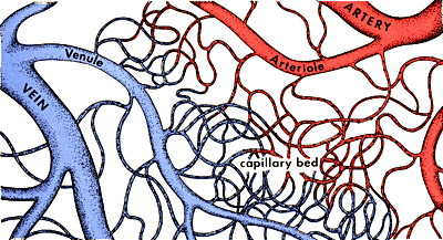
 3. Capillaries
Capillaries are extremely small vessels located within the tissues of the body that transport blood from the arteries to the veins. Capillary walls are thin and are composed of endothelium (a single layer of overlapping flat cells). Oxygen, carbon dioxide, nutrients and wastes are exchanged through the thin walls of the capillaries.
The flow of blood is controlled by structures called precapillary sphincters. These structures are located between arterioles and capillaries and contain muscle fibers that allow them to contract. When the sphincters are open, blood flows freely to the capillary beds of body tissue. When the sphincters are closed, blood is not allowed to flow through the capillary beds.
Capillaries are so small that red blood cells can only travel through them in single file.
- 5-10 microns in diameter.
 4. Sinusoids
The liver, spleen and bone marrow contain vessel structures called sinusoids instead of capillaries. Similar to capillaries sinusoids are composed of endothelium. The individual endothelial cells however do not overlap as in capillaries and are spread out. Oxygen, carbon dioxide, nutrients, proteins and wastes are exchanged through the thin walls of the sinusoids. The size of a sinusoid is around 30-40 microns in diameter.
additional information:
Microcirculation deals with the flow of blood from arterioles to capillaries or sinusoids to venules. Blood flows freely between an arteriole and a venule through a vessel channel called a thoroughfare channel. Capillaries extend from this channel and structures called precapillary sphincters control the flow of blood between the arteriole and capillaries.
The precapillary sphincters contain muscle fibers that allow them to contract. When the sphincters are open, blood flows freely to the capillary beds where gases and waste can be exchanged with body tissue. When the sphincters are closed, blood is not allowed to flow through the capillary beds and must flow directly from the arteriole to the venule through the thoroughfare channel.
It is important to note that blood is supplied to all parts of the body at all times but all capillary beds do not contain blood at all times. Blood is diverted to the parts of the body that need it most at a particular time. For instance when you eat a meal blood is diverted from other parts of your body to the digestive tract.
Vessel Sizes (Diameter in Microns)
Arterioles
20-50
Capillaries
5-10
Sinusoids
30-40
Venules
30-40
Images courtesy of:
MedValetCarolina Biological Supply/Access Excellence
transport system[2]
3:55 AM
In this post, the main focus will be on one of the main components of the circulatory system- the heart. Let's take a look on the anatomy of the human heart.
The heart sends blood around our body. The blood provides our body with the oxygen and nutrients it needs. It also carries away waste. Our heart is sort of like a pump, or two pumps in one. The right side of your heart receives blood from the body and pumps it to the lungs.
 Heart Parts
The heart is made up of four different blood-filled areas, and each of these areas is called a chamber. There are two chambers on each side of the heart. One chamber is on the top and one chamber is on the bottom. The two chambers on top are called the
atria. (singular: atrium) The atria are the chambers that are filled with blood returning to the heart from the body and lungs. The heart has a left atrium and a right atrium.
The two chambers on the bottom are called the
ventricles. The heart has a left ventricle and a right ventricle. Their job is to squirt out blood to the body and lungs. Running down the middle of the heart is a thick wall of muscle called the septum. The septum's job is to separate the left side and the right side of the heart.
The atria and ventricles work as a team— the right atrium pumps the blood to the right ventricle, which then pumps the blood to the lungs via the pulmonary artery. After passing through the richly vascularized lung tissue, oxygen-rich blood return to the heart via the pulmonary vein and into the left atrium. The left atrium pumps the blood to the left ventricle, the launching pad that pumps the blood through the huge aortic artery and delivers blood to the rest of the body.
Well, our blood relies on four special valves inside the heart. A
valve maintains the unidirectional flow of blood by opening and closing depending on the difference in pressure on each side. It keeps the flow of blood there by closing.
Two of the heart valves are the
mitral valve and the
tricuspid valve. They let blood flow from the atria to the ventricles. The other two are called the
aortic valve and
pulmonary valve, and they're in charge of controlling the flow as the blood leaves the heart. These valves all work to keep the blood flowing forward. They open up to let the blood move ahead, then they close quickly to keep the blood from flowing backward.
reference:
http://www.wanderings.net/http://en.wikipedia.org/wiki/Heart_valve
transport system[1]
6:51 AM
moving on from digestion, we will now move on to the next topic: transport in living thing. i am going to come up with one question to research on: what are the differences between the components of the transport system in humans and plants?
firstly, i will start off with human transport system. here is the main overview.
The circulatory system is an organ system that moves nutrients, gases, and wastes to and from cells, helps fight diseases and helps stabilize body temperature and pH to maintain homeostasis. The main components of the human circulatory system are the heart, the blood, and the blood vessels. The circulatory system includes: the pulmonary circulation and the systemic circulation.
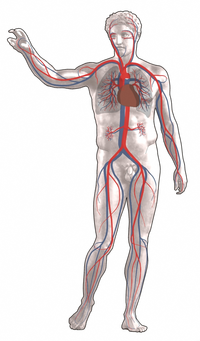 the human circulatory systemred indicates oxygenated bloodblue indicates deoxygenated blood
additional information:
pH: pH is the measure of the acidity or alkalinity of a solution.
Homeostasis: the property of either an open system or a closed system, especially a living organism, that regulates its internal environment so as to maintain a stable, constant condition.
you may not be able to understand some of the terms used here. but don't worry. in the next post, i will give you a more detailed explanation on each and every one of them(:
reference:
http://en.wikipedia.org/wiki/Homeostasishttp://en.wikipedia.org/wiki/PH
digestion[7]
5:43 AM
once food has passed through the small intestine, it is mostly undigestible material and water. it enters the large intestine, named for its wide diameter. the large intestine has six parts: the cecum, ascending colon, transverse colon, descending colon, sigmoid colon, and rectum.
the large pouch-shaped cecum marks the beginning of the colon. attached near the cecum bottom is the vermiform (worm-like) appendix. the appendix contains lymphoid tissue and intercepts pathogenic microorganisms that enter the digestive tract. sometimes, fecal matter may become trapped in the appendix, resulting in appendicitis (infection and inflammation).
the other parts of the colon absorb water and minerals from the undigested food and compact the remaining material into feaces. defecation is the digestive process final stage: feces (undigested waste products) are carried to the rectum through peristalsis and passed out through the anus.
additonal information:
vermiform appendix is a blind ended tube connected to the cecum, from which it develops embryologically. the cecum is a pouch-like structure of the colon. the appendix is near the junction of the small intestine and the large intestine. the appendix averages 10 cm in length and its diameter is usually between 7 and 8 mm.

the most common explanation is that the appendix is a vestigial structure with no absolute purpose. in The Story of Evolution, Joseph McCabe once argued:
the vermiform appendage—in which some recent medical writers have vainly endeavoured to find a utility—is the shrunken remainder of a large and normal intestine of a remote ancestor. this interpretation of it would stand even if it were found to have a certain use in the human body. vestigial organs are sometimes pressed into a secondary use when their original function has been lost.
given the appendix's propensity to cause death via infection, and the seemingly perfect health of those who have had their appendix removed, the biological purpose of the appendix has mystified scientists for some time. there have been cases of people who have been found, to have a congenital absence of an appendix. there have been no reports of impaired immune or gastrointestinal function in these people.
defecation is the final act of digestion by which organisms eliminate solid, semisolid or liquid waste material from the digestive tract via the anus. when humans expel feaces, waves of muscular contraction known as peristalsis in the walls of the colon move fecal matter through the digestive tract towards the rectum. undigested food may also be expelled this way. this process is called egestion.
information is taken from:
http://en.wikipedia.org/wiki/Vermiform_appendix
http://en.wikipedia.org/wiki/Defecation
digestion[6]
6:08 AM
now, i will continue from the previous post.
from the duodenum, chyme passes to the jejunum and ileum. here, tiny villi (finger-like projections) cover the walls of the small intestine. the cells that line the villi are covered with small projections called microvilli. these projections increase the surface area of the small intestine, allowing the chyme to contact more of the small intestine wall. the increased contact causes more efficient food absorption.
during food absorption, food molecules enter the bloodstream through the intestinal walls. capillaries within the villi absorb products of protein and carbohydrate digestion. lymph vessels (lacteals) within the villi absorb products of fat digestion and eventually lead to the bloodstream.
from the small intestine, digested products travel to the liver, one of the body's most versatile organs. hepatocytes (liver cells) detoxify blood of harmful substances such as alcohol and ammonia. And, hepatocytes store fat-soluble vitamins and excess substances such as glucose (sugar) for release when the body requires extra energy.
additional information about intestinal villi:
intestinal villi are tiny, finger-like projections that protrude from the wall of the small intestine and have additional extensions called microvilli which protrude from epithelial cells lining villi. they increase the absorptive area and the surface area of the intestinal wall.
it is important that the food is absorbed at a considerably fast rate so as to allow more food to be absorbed. (If the process is too slow, the concentration of the blood in the blood vessels and the food will be equal, thus, diffusion will not occur.) digested nutrients pass into the villi through diffusion. circulating blood then carries these nutrients away.

information is taken from:
digestion[5]
1:20 AM
The small intestine is about 20 feet (6 meters) long and has three parts: the duodenum, jejunum, and ileum. The duodenum is where most chemical digestion takes place. Here, bile from the gallbladder and enzymes from the pancreas and intestinal walls combine with the chyme to begin the final part of digestion.
Bile liquid is created in the liver and stored in the gallbladder. Bile emulsifies (breaks into small particles) lipids (fats), which helps in the mechanical digestion of fats. The pancreas and gland cells of the small intestine secrete digestive enzymes that chemically break down complex food molecules into simpler ones. These enzymes include trypsin (for protein digestion), amylase (for carbohydrate digestion), and lipase (for lipid digestion).
When food passes through the duodenum, digestion is complete.

in the next post, i will move on to the digestion in jejunum and ileum.
digestion[4]
9:43 PM
In the stomach, food undergoes chemical and mechanical digestion. Here, peristaltic contractions (mechanical digestion) churn the bolus, which mixes with strong digestive juices that the stomach lining cells secrete (chemical digestion).
The stomach walls contain three layers of smooth muscle arranged in longitudinal, circular, and oblique (diagonal) rows. These muscles allow the stomach to squeeze and churn the food during mechanical digestion.
Powerful hydrochloric acid in the stomach helps break down the bolus into a liquid called chyme. A thick mucus layer that lines the stomach walls prevents the stomach from digesting itself. When mucus is limited, an ulcer (erosion of tissue) may form.
Food is digested in the stomach for several hours. During this time, a stomach enzyme called pepsin breaks down most of the protein in the food. Next, the chyme is slowly transported from the pylorus (end portion of the stomach) through a sphincter and into the small intestine where further digestion and nutrient absorption occurs.
additional information:
chyme- a thick semifluid mass of partly digested food that is passed from the stomach into the duodenum, the first section of the small intestine.
pepsin- enzyme produced in the mucosal lining of the stomach that acts to degrade protein. Pepsin is one of three principal protein-degrading, or proteolytic enzymes in the digestive system. During the process of digestion, these enzymes, each of which is particularly effective in severing links between particular types of amino acids, collaborate to break down dietary proteins to their components, i.e., peptides and amino acids, which can be readily absorbed by the intestinal lining.
Pepsin is synthesized in an inactive form by the stomach lining; hydrochloric acid, also produced by the gastric mucosa, is necessary to convert the inactive enzyme and to maintain the optimum acidity (pH 1–3) for pepsin function. Pepsin and other proteolytic enzymes are used in the laboratory analysis of various proteins; pepsin is also used in the preparation of cheese and other protein-containing foods.
pepsin
information is taken from:
digestion[3]
8:23 PM
after the swallowing of the food, the next stop in the digestive system is the Esophagus, also known as the gullet.
the esophagus, a narrow, muscular tube about 30 centimeters long, starts at the pharynx, passes through the larynx and diaphragm, and ends at the cardiac orifice of the stomach. the wall of the esophagus is made up of two layers of smooth muscles. the inner layer of muscles is arranged circularly in a series of descending rings, while the outer layer is arranged longitudinally.
at the top of the esophagus, is a flap of tissue called the
epiglottis that closes during swallowing to prevent food from entering the trachea (windpipe). the chewed food is pushed down the esophagus to the stomach through
peristaltic contraction of these muscles. it takes only seconds for food to pass through the esophagus, and little digestion actually takes place.
additional information:
epiglottis- a lid-like flap of elastic cartilage tissue covered with a mucous membrane, attached to the root of the tongue. it is normally pointed upward, but during swallowing, elevation of the hyoid bone draws the larynx upward. as a result, the epiglottis folds down to a more horizontal position. in this manner it prevents food from going into the trachea and instead directs it to the esophagus.

peristaltic- the rhythmic contraction of smooth muscles to propel contents through the digestive tract.
After food is chewed into a bolus, it is swallowed to move it into the oesophagus. Smooth muscles will contract behind the bolus to prevent it from being squeezed back onto the mouth, then rhythmic, unidirectional waves of contractions will work to rapidly force the food into the stomach. This process works in one direction only and its sole purpose is to move food from the mouth into the stomach.
In the esophagus, two types of peristalsis occur:
first, there is a primary peristaltic wave which forces the bolus down the esophagus and into the stomach in a wave lasting about 8-9 seconds. The wave travels down to the stomach even if the bolus of food descends at a greater rate than the wave itself, and will continue even if for some reason the bolus gets stuck further up the esophagus.
In the event that the bolus gets stuck or moves slower than the primary peristaltic wave (as can happen when it is poorly lubricated), stretch receptors in the oesophageal lining are stimulated and a local reflex response causes a secondary peristaltic wave around the bolus, forcing it further down the esophagus, and these secondary waves will continue indefinitely until the bolus enters the stomach.

information is taken from:
http://en.wikipedia.org/wiki/Epiglottis
http://en.wikipedia.org/wiki/Peristalsis
digestion[2]
9:23 PM
the first stage of digestion begins in the mouth, where our teeth grind and chew food, breaking it into small manageable pieces. this chewing process, known as mastication, is dependent upon powerful muscles (masseter and temporalis), as well as smaller muscles that permit fine control. they move the lower jawbone against the upper jaw, enabling the crushing of hard food.
mastication causes exocrine glands under the tongue and in the back of the mouth to secrete a watery liquid called saliva which performs two essential functions:
1. it moistens and compacts the chewed food so the tongue can roll it into a ball (bolus) and push it to the back of your mouth for swallowing and easy passage through the pharynx and
esophagus.
2. saliva contains digestive enzymes which begin the breakdown of carbohydrates.
mastication and saliva secretion work in harmony, as chewing increases the surface area of food which helps to accelerate the breakdown of starch molecules into simple sugars by the digestive enzymes.
more facts about the salivary gland:
Salivary glands:
#1 is Parotid gland,
#2 is Submandibular gland,
#3 is Sublingual gland
the
parotid glands are a pair of glands located in the subcutaneous tissues of the face overlying the mandibular ramus and anterior and inferior to the external ear. the secretion produced by the parotid glands is serous in nature, and enters the oral cavity through the Stensen's duct. despite being the largest pair of glands, only approximately 25% of saliva is produced by the glands.
the
submandibular glands are a pair of glands located beneath the floor of the mouth, superior to the digastric muscles. the secretion produced is a mixture of both serous and mucous and enters the oral cavity via Wharton's ducts. approximately 70% of saliva in the oral cavity is produced by the submandibular glands.
the
sublingual glands are a pair of glands located beneath the floor of the mouth anterior to the submandibular glands. the secretion produced is mainly mucous in nature, however it is categorized as a mixed gland. approximately 5% of saliva entering the oral cavity come from these glands.
information is taken from:
http://en.wikipedia.org/wiki/Salivary_gland
digestion[1]
6:50 AM
to start off with the topic on digestion, i have come up with a question: what are the organs that make digestion possible in our bodies? it may seem as though we have already learnt it in primary school. but in fact, human digestion is much more than that and there are still many interesting facts and details to it, which i am going to cover later on.
a very simple definition about digestion is that it is the breaking down of food in the body, into a form that can be absorbed into the blood stream, and the remainders that cannot be digested will then be passed out of the body.
let's take a look about the whole of the digestive system:

my first entry
6:28 AM
my first entryhi all, this will be my first post in this biology learning journal. the two topics that i have decided to work on are:
1. digestion
2. transport in living things
ok, that is all i have for now. the research that i have done for the two topics will be put on the next post. bye(:


